Photos of AFB
This image illustrates the contents of an AFB infected cell that is at the midpoint between a 'wet' ropable larvae and a dry black scale. Of note is the darker brown colour due to age.
This image illustrates the contents of an AFB infected cell that is at the midpoint between a 'wet' ropable larvae and a dry black scale. Of note is the darker brown colour due to age, the glossy appearance and the more gelatinous nature of the cell contents. Additionally, the volume of the cell contents is substantially less. Photo supplied by Murray Rixon
This image illustrates the coffee colour development in two larvae. Note also the horizontal lines sometimes seen that is referred to as 'loss of body segmentation'. Photo supplied by Murray Rixon
AFB infected larvae at pupal stage - Bob Russell
Decomposing pupa. In bright sunlight, the head and body appear very pale. The mouth and tongue can be seen clearly. Photo supplied by Murray Rixon
slumped coffee coloured larvae. Note the shiny appearance, the even tone of colour and the way a slumping larvae appears to rise up at the back.
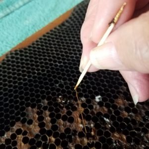
Roping out - photo by Tim Sharp
Two decomposing larvae. The brown/black prepupal heads are foreground in the cells. Note the distinct coffee colour in comparison to healthy eggs and larvae. Photo supplied by Murray Rixon
Partial cell capping removal and from the dark within, mouth and tongue in early development.
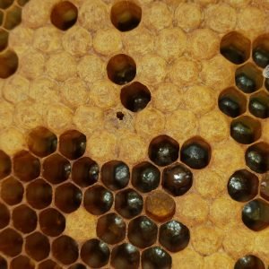
Four AFB-infected cells in a hive. Larval colour darkens with time, contents become tacky, and material lessens. Blackened, sunken, and perforated cappings are also visible.
This image shows five AFB-infected larvae and pupae. It highlights three slumping stages, two pupal infection stages with visible mouthparts and tongue, and desiccation shrinkage. Photo: Murray Rixon
This image shows the colour, texture, and volume of a long-term AFB-infected cell. The roping length has reduced, and little material remains on the matchstick. Photo: Murray Rixon.
This image shows a desiccated AFB variation with a dry scale body and dried pupal tongue. The black, glossy scale has creased edges due to desiccation. Photo: Murray Rixon.
This photo shows four slumped larvae with a concave shape (three below center, one top right). Three perforated cappings at the top vary from pinholes to fully exposed cells. Photo: Murray Rixon.
Pupa tongue
This image shows a larva decomposing at the pupal stage. It has a uniform coffee colour, lost segmentation, and is just starting to slump into a concave state. Photo: Murray Rixon.
This image shows a slumping larva with faint horizontal lines and a raised rear. Its pale colour indicates an early stage, photographed on a sunny day. Photo: Murray Rixon.
Photo shows AFB slumped in a cell surrounded by eggs. Bees have removed 90% of the cell cap but haven’t yet cleared the infected contents. Photo: Murray Rixon.
This image shows two decomposing pupa side by side. Note the slight colour variation and shape difference with the two pupa. Photo supplied by Murray Rixon
This image shows AFB in older frames. An infected pupa with pupal tongue is found away from the brood, in crystallized honey. Matchstick shows size. Photo: Murray Rixon.
This photo shows five desiccated shiny black scales. The scales adhere firmly to the cell floor and can't be pulled out with a matchstick. Photo: Murray Rixon.
This heavily infected AFB frame shows darkened, greasy, and sunken caps. Perforations range from tiny pinholes to large chewed caps. Photo: Murray Rixon.
This image shows a common response to stirring an AFB-infected cell. A bee begins licking the material. The cell will be empty within hours. Photo: Murray Rixon.
Pupa tongue
This image shows four cells with clear signs of AFB. Photo supplied by Murray Rixon
This photo shows an unusual case: two bee pupae in one cell, both collapsed with AFB and displaying pupal tongues. Photo: Murray Rixon.
This image shows two AFB-infected cells in an interim stage between coffee-coloured and desiccated black scale. Note the uniform colour, texture, and glossy appearance. Photo: Murray Rixon.
Frame with AFB, photo courtesy of Kintail
Uncapping a sunken perforated cell exposed an AFB-infected larva that died at pupal stage. Note the infected cell surrounded by fresh egg cells. Photo: Jason Ward.
Burning AFB infected hive, photo courtesy of John Graham
This photo shows two views of AFB: left, late onset with coffee colour and pupal tongue; right, slumped AFB larva with lost segmentation. Photo: Murray Rixon.
After uncapping sunken perforated cells, AFB larvae were found. Top: AFB larva died at pupal stage, bottom: AFB larva at pre-pupal stage. Photo: Jason Ward.
AFB killed larva at pupal stage AFB, courtesy of Kintail
Scale - photo courtesy of Bob Russell
This photo shows AFB roping out to 30mm. Note the consistent colour and texture with no lumps or watery areas, just before snapping off. Photo: Murray Rixon.
This photo shows AFB material roping out under UV light, just before separating. It’s even in consistency with no lumps or inclusions. Photo: Murray Rixon.
This photo shows AFB with a spotty brood pattern. Note the perforated cappings, including one cell where AFB is visible inside. Photo: Murray Rixon.
This photo shows a slumped AFB larva with lost body segments and horizontal lines. Bees removed 90% of the capping but left the infected material. Photo: Murray Rixon.
Photo illustrates the coffee coloured AFB. Note the loss of body segments - Murray Rixon
Greasy sunken perforated cells, pupal tongue and pre-pupal AFB scale in dead out hive - photo courtesy of Lott Larson
Photo illustrates AFB scale. It is shiny black, hardened, difficult to remove. Will fluoresce under UV light - Murray Rixon
Burning AFB infected hives - Murray Rixon
Rare finding two AFB pupal tongue in the same cell. Photo supplied by Matt Tubbs.
Burning of AFB infected hive in backyard, photo courtesy of Chris Crook
Making a killed AFB positive hive bee proof and preparing it for safe transport to the burning pit, photo courtesy of Chris Crook
AFB ropiness test on newly killed AFB affected larva photo courtesy of Chris Crook
Infected AFB larva that died at the pupal stage. The white margins of the body are still clearly visible. Photo courtesy of Murray Rixon
AFB Pupa scale photo courtesy of Bob Russell
Pupa tongue courtesy of Bob Russell
Roping out - photo courtesy of Bob Russell
Roping out - note the consistent colour and texture. It is shiny and there are no lumps or solids - Murray Rixon
AFB infected brood cells cappings at early stages don't necessarily show very different colour from healthy capped brood
Diseased pupa with tongue
AFB Roping Out
AFB
photo gallery
We invite all beekeepers to send in their photos of AFB and the destruction of their hives in order to help educate other beekeepers. Please send your photos to info@nzbb.org.nz with a brief description of each shot. For best results, the photos need to be in high resolution.